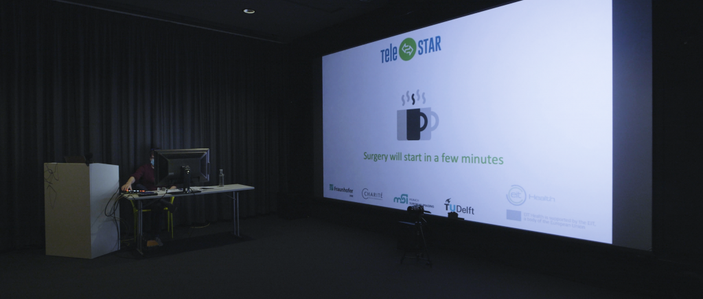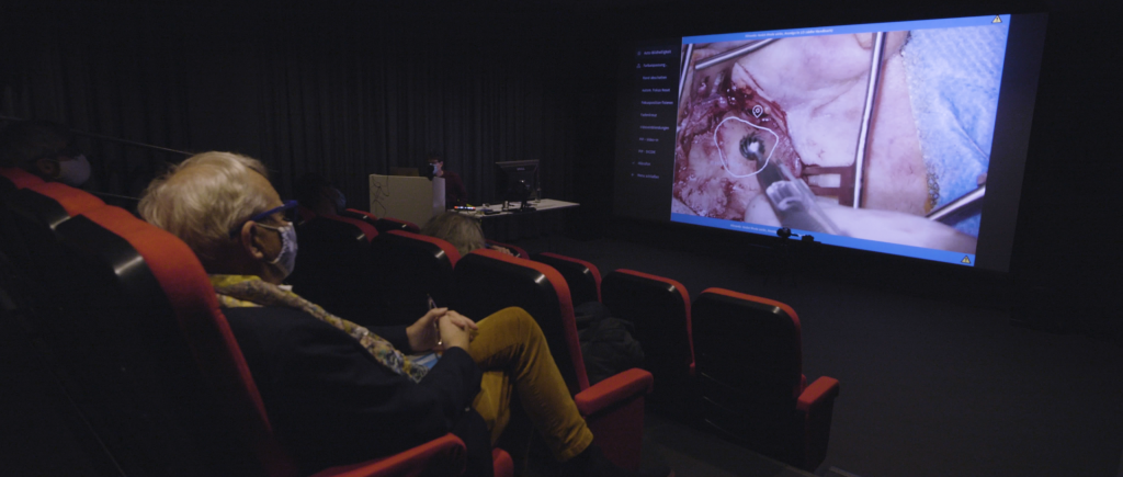

Berlin, 15 November 2020 – For the second time, 3IT’s partner project TeleSTAR successfully transmitted the surgery of a cochlear implantation using an augmented reality based 3D live stream from ENT clinics of Berlin’s renowned hospital university Charité. This time, the stream was broadcasted to four lecture halls Europe-wide, hence, it permitted 45 viewers to witness the surgery live. Students from the ENT department of the hospital ErasmusMC in Rotterdam, one of the most prestigious research hospitals in Europe, as well as from the living lab of trauma surgery in the University hospital of Ludwig-Maximiliam University in Munich were able to follow the surgery for teaching purposes live with a latency of 800ms. Moreover, other surgeons from the field of visceral and transplant surgery as well as medical master students could survey the surgery in 3IT’s 3D-cinema, equally with a latency of 800ms. The additional broadcast to Charité’s Intranet gave students the advantage to observe the surgery live with a latency of mere 20ms. Subsequent to the live streaming, 67 viewers watched the 2D YouTube stream, with a latency of 8s.
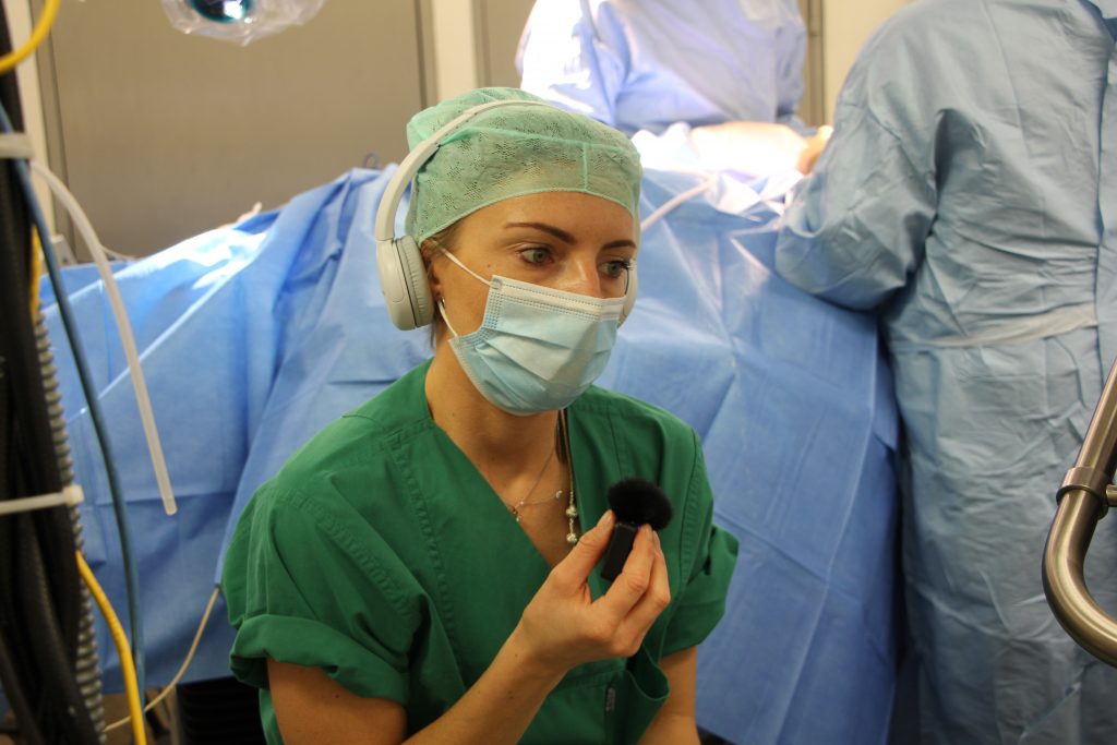
This time, an improved audio concept helped to relief the operating senior surgeon, Dr. Florian Uecker. Once again, the 3D stream was supported by synchronous audio commentary and an intraoperative annotation mode using AR. Thus, every single step was conveyed by explanations, making them more comprehensible for the students. By means of the intraoperative annotation mode, surgeons were able to append visual information to the live stream, in the form of sketches, references and anatomy measurements. Even more, the students had the opportunity to ask questions from the various remote lecture halls to the second assistant surgeon Dr. Sophia Häusler.
As the OR is a time and resource constrained environment, live surgeries enjoy a long tradition within medical education, already. However, the integration of innovative AR approaches, facilitates groundbreaking new teaching methods. The live transfer did not solely enable a larger public to monitor the surgery live. In fact, in times of Covid19, this new format of teaching becomes pivotal, rendering possible high quality distant learning without putting anybody at unnecessary risk.
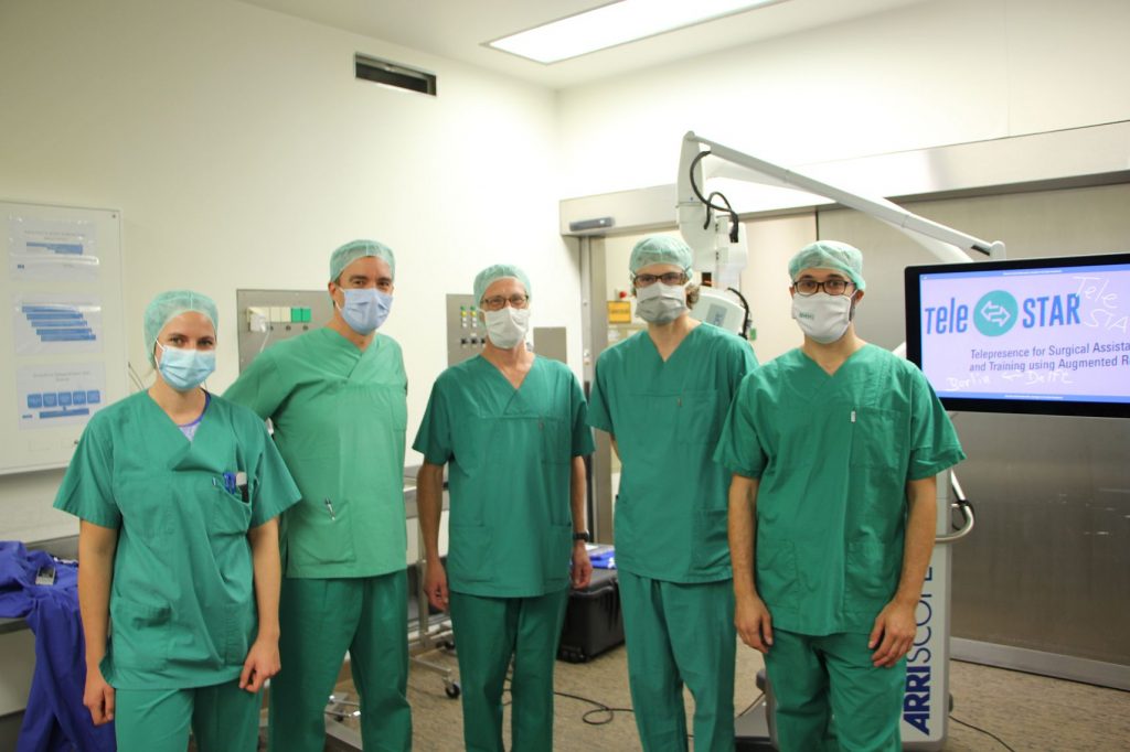
The insertion of a cochlear implant is a standard operation in ENT surgery, in order to treat deaf people. The implementation is advantageous in order to demonstrate the use of AR-technology and to test it, as the surgery can be made within a short time. The fully digital surgical microscope “ARRISCOPE” of the company Munich Surgical Imaging (formerly ARRI Medical) was used for the visualizations, as it renders possible high-resolution 3D images, a radiation-free and image-based measurement of the anatomy and the acquisition of multispectral image data.
TeleSTAR is funded by the EIT-Health. EIT-Health is supported by the EIT, a body of the European Union.
Find all information on our 3IT-Project TeleStar here.
Find the press release to the 3D live surgery here.
Expert opinions
Dr. R.M. (Mick) Metselaar Mick, ENT surgeon at Erasmus MC:
“I’m definitely hoping for a follow-up.”
Prof. Dr. Reiner Kunz, visceral surgeon, Berlin-Tempelhof:
“Congratulations on this absolutely impressive demonstration of the Arriscope System on the occasion of cochlear implantation. In my opinion, extremely good image, excellent 3-dimensionality and the additional attributes such as ROI markings, tissue recognition etc. were demonstrated very effectively and in my opinion already very well suited for routine operation.”
Prof. Dr. John v.d. Dobbelsteen, faculty of Biomedical Engineering at TU Delft:
“Students and clinical trainees need to experience the full spectrum of modern medical technology. TeleSTAR offers surgical trainees and medical technology developers this important exposure”
N.N., Biomedical Engineer, TU Delft
“Amazing experience to observe a surgery like this. I found it very useful to be able to ask the surgeon exactly where we should be focusing on, this was only further helped by the ability for the surgeons to point out exact locations using the annotation mode.”
Dr. med. Florian C. Uecker, leading senior physician, ENT clinic at Charité Berlin, Virchow-Campus
“This technology provides an unparalleled quality of remote teaching and communication, which is particularly valuable given the current COVID-19 pandemic”
Dr. med. Steffen Dommerich, leading senior physician, ENT clinic at Charité ,Campus Mitte
“A surgical broadcast with all this additional information makes the whole thing appear much more descriptive and increases the learning experience.”^
Dr. med. Philipp Arens, ENT senior physician, ENT clinic at Charité, Campus Mitte
“A fascinating system, which gives a deep insight into the patient that is almost the same as if you were operating yourself”.
Dr. PM Ute Morgenstern, Biomedical Engineer, TU Dresden:
“This is really a very good way to provide a realistic environment for medical students far away from the surgical location, especially if the analogue training possibilities are now limited due to Corona. Above all – what interests us as biomedical engineers – the possibility to convey technical information about the medical environment in such a way that engineering students or practitioners can see exactly the interface between their research/development and the clinical user and can therefore approach their own work with more knowledge, motivation and criticism.”